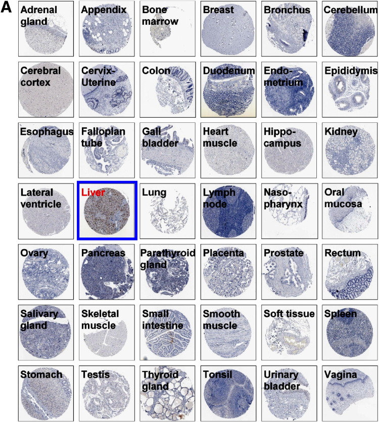
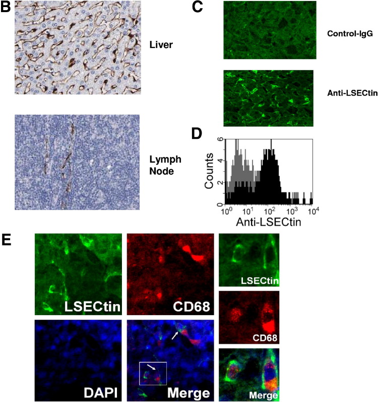
Supplementary Figure 1.
LSECtin specifically expresses in LSECs and Kupffer cells (A) Tissue array of LSECtin expression in 42 normal human tissues. All immunohistochemically stained sections were scanned using an automated slide-scanning system at 20× magnification. (B) Highlight of LSECtin expression in liver or lymph node from tissue array. (C) Immunofluorescein staining of normal liver sections was processed using antibody against LSECtin. (D) Expression of LSECtin in LSECs. Purified LSECs were stained with anti-LSECtin mAb. (E) Expression of LSECtin in Kupffers. Normal liver sections were stained with the antibodies against LSECtin (green) and CD68 (red).


