
Figure 1.
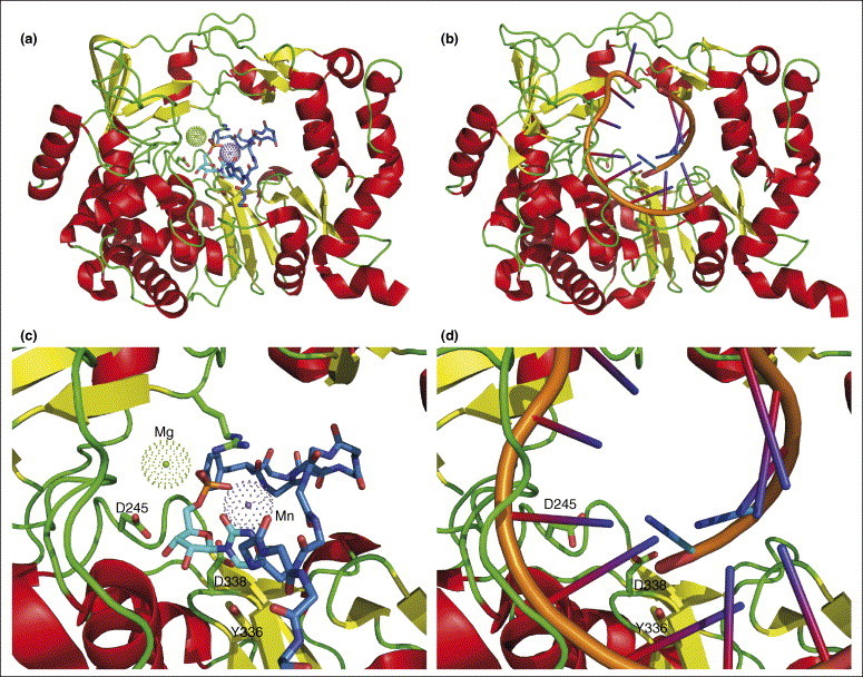
Comparison of polymerase-bound protein and RNA primers. VPg–UMP, overview in (a) and details in (c), is observed in the crystal structure ([22••]; PDB code 2F8E) in a position remarkably similar to that of the template–primer RNA duplex, overview in (b) and details in (d) ([23]; PDB code 1WNE). Except for Tyr3, only the mainchain atoms of VPg are shown for clarity: carbon atoms of VPg are colored blue and those of UMP cyan. Magnesium and manganese ions are shown as dotted balls in (a and c) and the template–primer RNA duplex is in stick form in (b and d). As a point of reference, three amino acids (of conserved elements in the active site) are shown as stick models: D245, Y336 and D338.