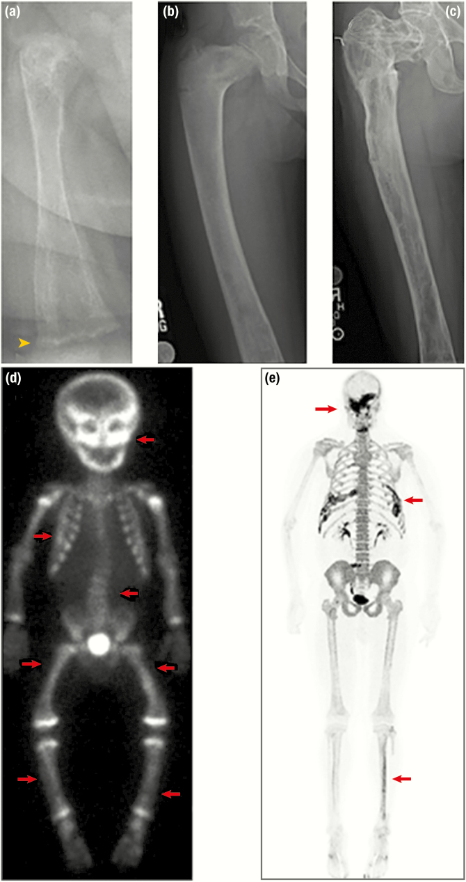
Figure 7.
Radiographic features of fibrous dysplasia (FD). A, B, and C, Upper panels demonstrate age-related radiographic changes in 3 patients with diffuse femoral involvement. A, In a 6-month-old patient, FD appears heterogeneous with streak-like features. Note the irregular metaphyses resulting from uncontrolled FGF23-mediated hypophosphatemia (yellow arrowhead). B, Radiograph from a 6-year-old demonstrates the classic “ground-glass” homogeneity. C, In a 31-year-old, FD again appears heterogeneous, with sclerotic areas interspersed with areas of radiolucency. D and E, Lower panels depict nuclear medicine scan images used to evaluate total skeletal FD burden. D, Technetium-99 scintigraphy scan in a young child with near panostotic disease shows increased tracer uptake in most of the skeleton, including the skull, spine, and long bones (red arrows). Note the symmetric increased uptake at the metaphyses in this growing child. E, 18F-NaF PET/CT scan in an adult with mild disease shows tracer uptake in areas of FD involving the skull, ribs, and left tibia (red arrows). Note the superior resolution and anatomical characterization of 18F-NaF in comparison to technetium.

