
Fig. 1.
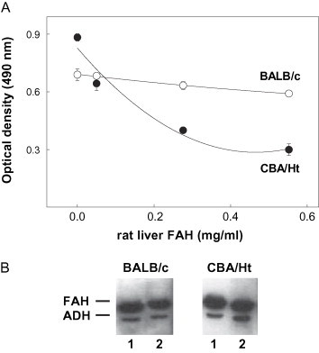
Reactivity of soluble rat liver FAH with the autoAb induced in MHV-infected mice. (A) Competition ELISA assays. Plates coated with rat liver FAH were incubated with 1:100 diluted serum from MHV-infected mice in the presence of different concentrations of the soluble enzyme. Bound Ab were revealed with peroxidase-labeled IgG anti-mouse IgG and OPD. (B) Western-blot competition assays. Rat liver FAH (1 μg) was run in 10% SDS-PAGE, transferred onto nitrocellulose sheets and incubated with a 1:200 diluted serum from MHV-infected mice in the absence (lane 1) or in the presence (lane 2) of soluble FAH (100 μg/ml). Densitometric values, expressed as percentage of intensity in lane 1, were 90% and 99% for sera from BALB/c and CBA/Ht mice, respectively. Results presented in the figure were obtained with sera from a BALB/c or a CBA/Ht mouse 30 days post MHV-infection.