
Fig. 6.
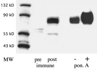
Western blot analysis of GP73 expression in HepSK-1 and GP73-transfected 293 cells. Cellular proteins were extracted from cultured HepSK-1 and GP73-transfected 293 cells, separated by SDS–PAGE (15 μg/lane), transferred to a PVDF membrane, and probed with a polyclonal rabbit antibody (dilution 1:5000) raised against GP73 (aa 41–400). The positions of molecular weight (MW) markers are indicated. Pre-, post-immune: lysates of HepSK-1 cells were probed with prei-mmune sera or post-immune sera from week 8 after injection respectively. PonA: 293 cells were incubated in the absence (−) or presence (+) of 5 μM ponasterone A for 16 h, and cellular lysates were probed with post-immune sera. Signals were detected using an HRP-conjugated secondary antibody (1:1500), and visualized by ECL. The immune sera specifically recognize a broad protein band of approximately 73 kDa (GP73). GP73 expression is strongly induced after ponasterone A induction in 293 cells.