
Fig. 1.
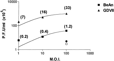
Semilog graph showing virus production in supernatants of BeAn- and GDVII virus-infected astrocytes measured by plaque assay on BHK-21 cell monolayers. Cells (3 × 10³) were infected at m.o.i.s of 1, 10, and 100 for 45 min at room temperature. Residual virus remaining from the inoculum were washed three times and cultures were replenished with complete medium. The supernatants were centrifuged and tested 24 h postinfection. Circles (•, ○) indicate titers detected at a m.o.i. of 100 just after the washes were completed and numbers in parentheses indicate PFU/cell.