
Figure 4.
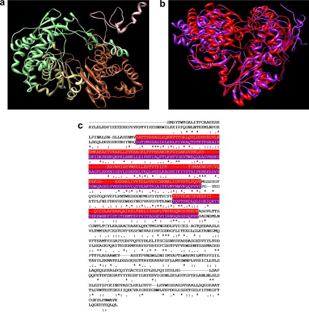
(a) Overview of the entire structure of the RdRp of BVDV. The protein domains are colored as follows: pink, N-terminal domain (residues 71–138); light green, fingers domain (residues 139–313 and 351–410); palm domain, kaki (residues 314–350 and 411–500); sienna, thumb domain (residues 501–679). (b) Overlay of the 3D models of RdRp of BVDV (purple) and HCV (red). (c) BVDV and HCV polymerase amino acid alignment: top line, BVDV (residues 92–679); bottom line, HCV (residues 1–531). The alignment of the fingers domain is highlighted in purple (BVDV) and red (HCV).