
Fig. 3.
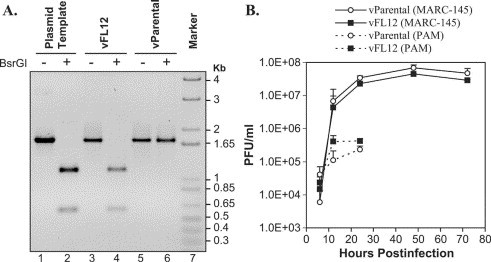
Characterization of rescued virus. (A) An amplicon of 1740 bp, spanning the genetic marker, was amplified by RT-PCR using total RNA from cells infected with either vFL12 (lanes 3 and 4) or parental virus (lanes 5 and 6). pFL12 DNA also was used in PCR amplification as a positive control (lanes 1 and 2). The amplicons were digested with BsrGI. DNA was electrophoresed on 1% agarose gel and an inverted image was acquired. (B) Growth kinetics of cloned and parental virus. MARC-145 cells (2.5 × 10⁶) were infected with either vFL12 or parental virus. At 6, 12, 24, 48, and 72 hpi, supernatant was collected and titrated by plaque assay. PAMs were infected and cultures were collected at 6, 12, and 24 hpi for titration of infectivity. Error bars represent standard deviation from replica experiments.