
Figure 3.
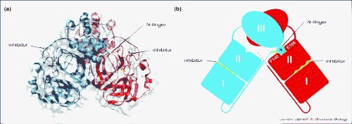
Nsp5, the SARS-CoV Mpro. (a) The crystal structure of SARS-CoV Mpro in complex with a CMK inhibitor (PDB code 1UKW). Protomers A and B are shown in ribbon representation, and are coloured red and blue, respectively. The CMK inhibitors are shown in yellow stick representation. The N-finger, residues 1–7 of protomer B, is shown in green. A transparent molecular surface is shown covering the structure. (b) Schematic of the SARS-CoV Mpro dimer, corresponding to the view in (a). Residue S1 on the N-finger of protomer B forms hydrogen bonds with two residues in protomer A, F140 and E166.