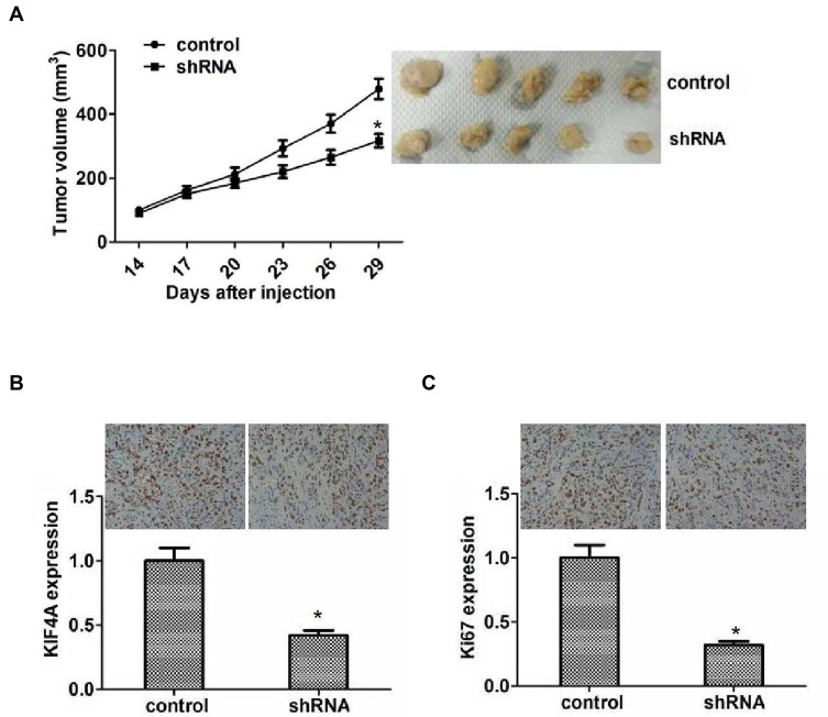
Figure 5.
KIF4A facilitated ccRCC growth in mice. (A) Hep3B cells infected with control or KIF4A shRNA lentivirus were subcutaneously implanted into nude mice. After 2 weeks, tumors were isolated, and volume was examined every 3 days (n=5 in each group). Tumor growth curve was calculated and analyzed according to the average volume of six tumors in each group. (B) IHC assays indicated the expression level of KIF4A in control or KIF4A depletion tumor tissues isolated from mice. (C) IHC assays revealed the expression level of Ki67 in control or KIF4A depletion tumor tissues taken from mice. Results are presented as mean ± SD, *P < 0.05.

