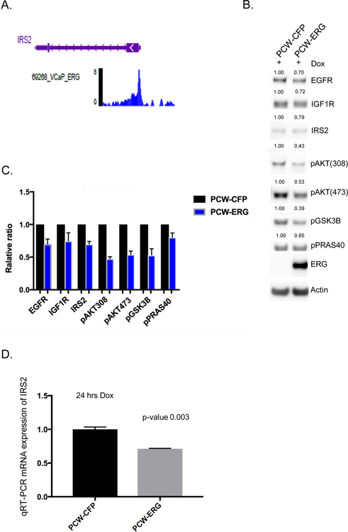
Figure 4. Validation of ERG repression of IRS2-RTK signaling in human model systems.
A) In silico ChIP seq analyses in the prostate cancer ERG positive cell line VCaP demonstrates significant enrichment for ERG binding at the IRS2 promoter region. Representative read peaks of ERG binding in the IRS2 transcription start site region. B) Dox-inducible overexpression of ERG (24 hrs) in human normal prostate organoids demonstrates reduced levels of IRS2, total EGFR, total IGF1R, and downstream PI3K phosphorylation of AKT, GSK3B, PRAS40. Experiment performed in triplicate, representative western blot shown, and quantification of proteins normalized to actin. C) Bar graph representing mean and standard deviation for protein quantification across three independent experiments of ERG over-expression in human normal prostate organoids, normalized to control. D) IRS2 mRNA levels are significantly repressed following acute overexpression of ERG (24 hrs) in human normal prostate, experiment performed in triplicate, and mean and standard deviation reported.

