
Figure 3. Elevated OGT and O-GlcNAc increases mammosphere formation and CD44HCD24L TIC population.
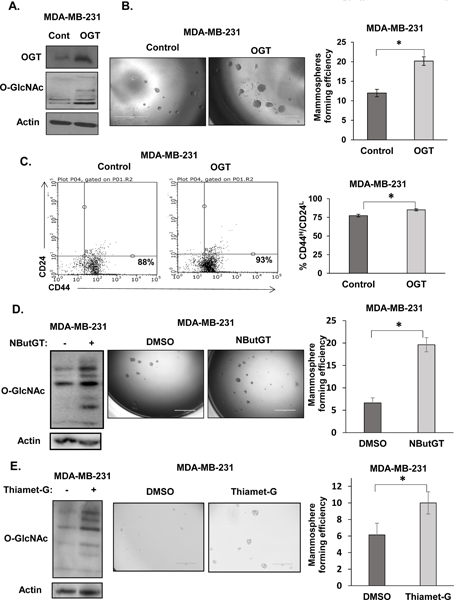
A. Cell lysates from MDA-MB-231 cells stably overexpressing control or OGT were collected for immunoblot analysis with indicated antibodies. B. Representative images of mammospheres formation of MDA-MB-231 cells stably overexpressing control or OGT (left) and quantification of MFE (right). C. Representative flow cytometry measuring CD44HCD24L population from MDA-MB-231 cells stably overexpressing control or OGT (left) and quantification (right). D. Cell lysates from MDA-MB-231 cells treated with DMSO and OGA inhibitor NButGT (100 μM) were collected for immunoblot analysis with indicated antibodies (left), representative mammospheres formation (center) and quantified MFE following 5–7day mammosphere culture (right). E. Cell lysates from MDA-MB-231 cells treated with DMSO and OGA inhibitor Thiamet-G (1 μM) were collected for immunoblot analysis with indicated antibodies (left), representative mammospheres formation (center) and quantified MFE following 7 day mammosphere culture (right). Student’s t-test reported as mean ± SEM. * = p-value < 0.05.