
Figure 1. P-AscH− increases ROS production 48 h after exposure, resulting in clonogenic cell death.
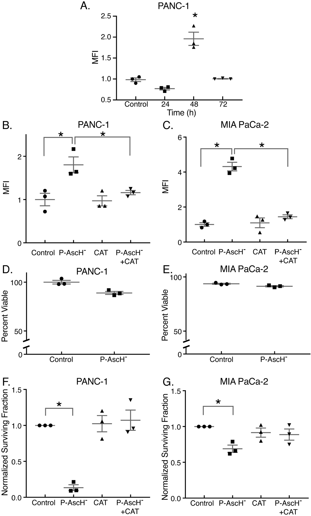
PDAC cell lines were treated with P-AscH− for 1 h for all experiments. Assays were performed 48 h after exposure, unless stated otherwise. Presented are means ± SEM for each set, n = 3; *p < 0.05 vs. control for all experiments. For panels A - C and F - G, ANOVA with Tukey’s multiple comparisons were used. For panels D - E, 2-tailed student’s t-test was used.
A. Time course (24 – 72 h) for the oxidation of DCFH-DA in PANC-1 cells treated with 2 mM ascorbate. Data is represented as mean fluorescent intensity (MFI) that is normalized to control.
B-C. PANC-1 and MIA PaCa-2 cells treated with P-AscH− (2 mM or 1 mM, respectively) exhibit increased DCFH-DA oxidation 48 h after treatment that is rescued by 100 μg/mL of bovine catalase in the medium.
D-E. PANC-1 and MIA PaCa-2 cells treated with 2 mM or 1 mM ascorbate respectively, exhibited no changes in viability using the propidium iodide (PI) exclusion assay (p > 0.05).
F-G. PANC-1 and MIA PaCa-2 cells treated with P-AscH− (2 mM or 1 mM, respectively) exhibit significant decreases in clonogenic survival.