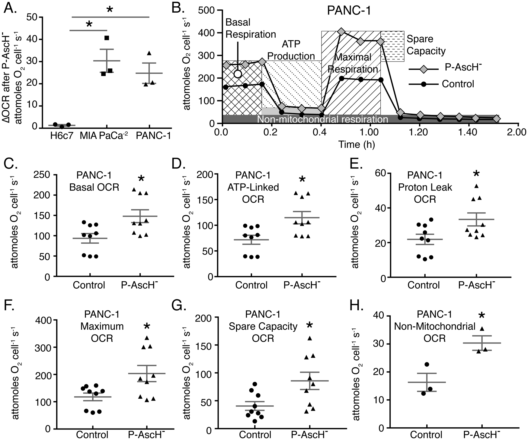
Figure 2. P-AscH− treatment results in increased basal oxygen consumption rate, ATP linked, Maximal Respiration, and Proton leak in PDAC cell lines.

A. PANC-1, MIA PaCa-2, and H6c7 cells were treated with P-AscH− for 1 h and basal oxygen consumption rate (OCR) was measured 48 h after treatment using a Clarke Electrode. Changes in OCR after P-AscH− treatment were determined by subtracting the untreated basal OCR from the increase following P-AscH− treatment to get ΔOCR. PANC-1 cells treated with 2 mM P-AscH−, MIA PaCa-2 cells treated with 1 mM P-AscH− and H6c7 cells treated with 2 mM P-AscH− (Means ± SEM, n = 3, *p < 0.05 vs. control, ANOVA with Tukey’s multiple comparisons).
B. Example data from Seahorse XF96 instrumentation showing that PANC-1 cells treated with P-AscH− (2 mM) have alterations in the mitochondrial stress test curves 48 h after exposure.
C-H. PANC-1 cells demonstrate an increase in: basal respiration; D. ATP production; E. proton leak; F. maximal respiration; G. spare capacity and H. non-mitochondrial respiration 48 h after treatment (Means ± SEM, n = 9 C-G and n = 3 H, p < 0.05 vs. control, 2-tailed student’s t-test).
