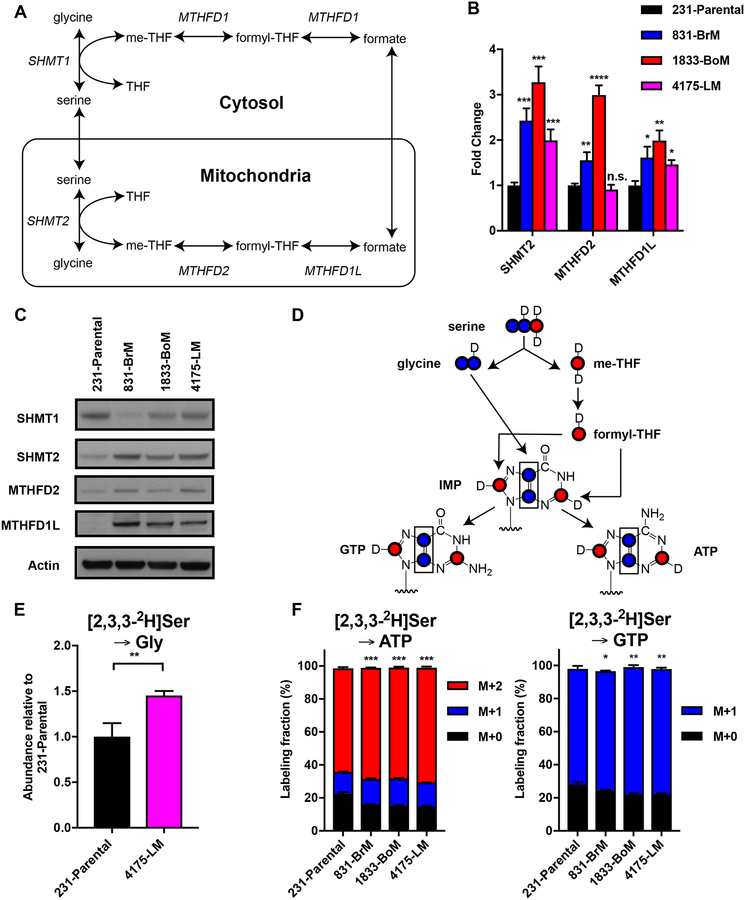
Figure 3.
The mitochondrial serine and one-carbon unit pathway is upregulated in metastatic breast cancer subclones. (A) Schematic of the cytosolic and mitochondrial serine and one-carbon unit pathway. (B) qPCR for serine and one-carbon unit pathway genes (mean ± SD, n = 3, *P < 0.05 **P < 0.01 ***P < 0.001 ****P < 0.0001 by two-tailed Student’s t test, compared to expression in parental cells). (C) IB for serine and one-carbon unit pathway enzymes from whole-cell extracts of parental cells and metastatic subclones. (D) Schematic diagram of incorporation of 2H (D) from [2,3,3-2H]serine onto glycine, one-carbon units, and purines. (E) SHMT flux estimated by relative abundance of labeled glycine from serine (mean ± SD, n = 3, **P < 0.01 by two-tailed Student’s t test). (F) Fractional labeling of [2,3,3-2H]serine onto GTP and ATP (mean ± SD, n = 3, *P < 0.05 **P < 0.01 ***P < 0.001 by two-tailed Student’s t test).

