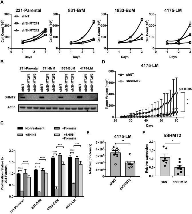
Figure 4.
Metastatic subclones are particularly sensitive to SHMT2 inhibition. (A) 3 day proliferation of 231-Parental, 831-BrM,1833-BoM, and 4175-LM cells expressing either a nontargeting (shNT) or SHMT2 targeting (shSHMT2) vectors. Relative proliferation was calculated relative to average proliferation of shNT cells (mean ± SD, n = 3). (B) IB for SHMT2 in parental and metastatic subclones. (C) 3 day proliferation of parental and metastatic cells with 2 μM SHIN1, in RPMI with or without 2 mM formate and dialyzed FBS (mean ± SD, n = 3, ***P < 0.001 ****P < 0.0001 by two-tailed Student’s t test). Counts were normalized to the proliferation of 231-Parental cells in media without SHIN1 and formate treatment. (D) Growth of 4175-LM shNT and shSHMT2 tumors in the mammary fat pad of nude mice (mean ± SEM, n = 8, **P < 0.01 by two-tailed Student’s t test). (E) Quantification of luminescence signal in the lungs of mice 3 weeks post injection of either 4175-LM shNT or shSHMT2 cells (mean ± SEM, **P < 0.01 by two-tailed Student’s t test, shNT;n = 8 shSHMT2;n = 7). (F) qPCR analysis of hGAPDH expression in the lungs of mice 4 weeks post injection of either 4175-LM shNT or shSHMT2 cells (mean ± SEM, *P < 0.05 by two-tailed Student’s t test, shNT;n = 6 shSHMT2;n = 7).

