Figure 1.

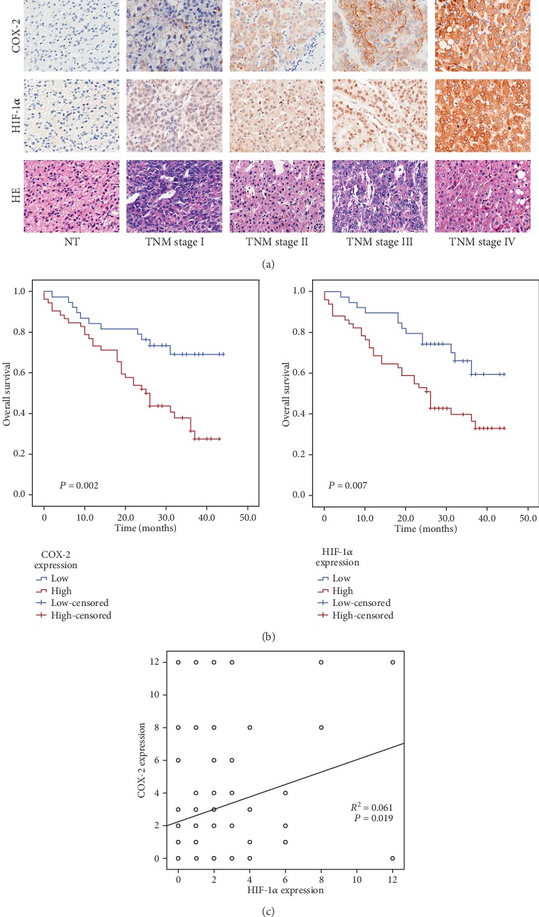
COX-2 and HIF-1α expressions in HCC tissues. (a) IHC staining of COX-2 and HIF-1α and HE in HCC tissue (magnification ×200). (b) High COX-2 or HIF-1α expressions correlate with poorer overall survival (P = 0.002 and P = 0.007, respectively, log-rank test). (c) Positive correlation between COX-2 expression and HIF-1α level (Pearson's correlation, R2 = 0.061, P = 0.019).
