
Fig. 1.
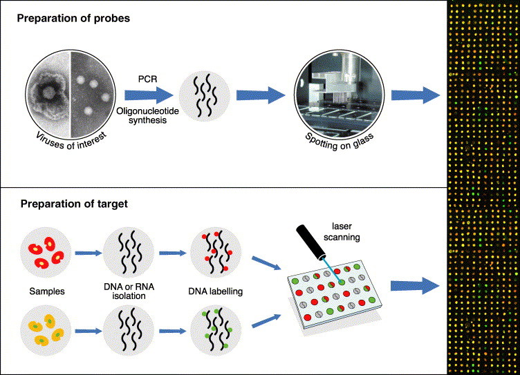
An illustration of the processes involved in making and using an array. At the top is depicted the route from the virus of interest to the spotted arrays (herpesvirus and enterovirus particles are shown). At the bottom, DNA or RNA is extracted from samples, amplified and labelled with either Cy3-dCTP (green) or Cy5-dCTP (red). When applied to the array bearing the immobilised probes, the target binds to complementary sequences. An example of an array result is shown on the right: the green spots represent hybridisation of the probe only with target sequences labelled with Cy3-dCTP; the red spots represent hybridisation of the probe only with target sequences labelled with Cy5-dCTP; the yellow spots represent hybridisation with both target sequences.