
Fig. 1.
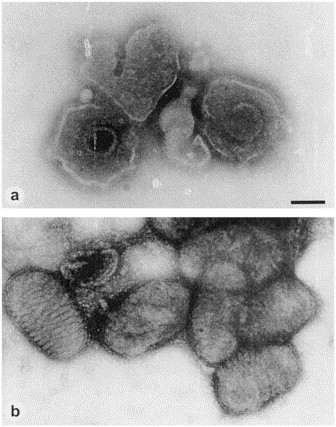
Direct EM of (a) herpesvirus particles from a varicella vesicle and (b) parapoxvirus recovered from the skin ulcers of a diseased seal, demonstrating the principles of morphological diagnosis after negative staining and the advantages of using different stains. The membrane destroying and/or penetrating effect of 2% phosphotungstic acid helps to reveal the 100 nm herpesvirus capsid within the viral envelope. 1% uranyl acetate on the other hand, by its remarkable membrane stabilizing effects, reveals the surface detail of the brick-shaped poxvirus very clearly. Magnification, ×80 000; bar, 100 nm.