
Fig. 3.
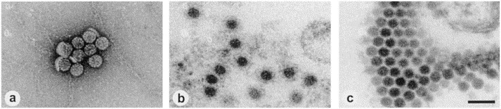
Human polyomavirus from the urine of bone marrow transplant recipients. (a) Negative staining after ultracentrifuge enrichment (100 000 ×g, 1 h, 20°C) using 1% uranyl acetate. (b) Thin section EM of a sediment (see a) embedded in Epon showing an immune aggregate. (c) Thin section EM of diagnostic HEL cell cultures inoculated with urine after ultracentrifuge enrichment. A paracrystalline array of polyoma virus particles is displayed. Magnification, ×80 000; bar, 100 nm.