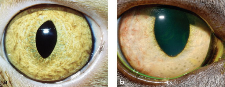
Fig 3 (a).

The normal anterior iris face is highly textured when viewed with oblique illumination and a source of magnification. (b) With uveitis, iridal swelling is evident as a ‘muddy’ or flattened iris surface, sometimes in association with nodular swellings. This eye also demonstrates rubeosis iridis (especially laterally) and keratic precipitates as grey spots against the inner cornea ventromedially
