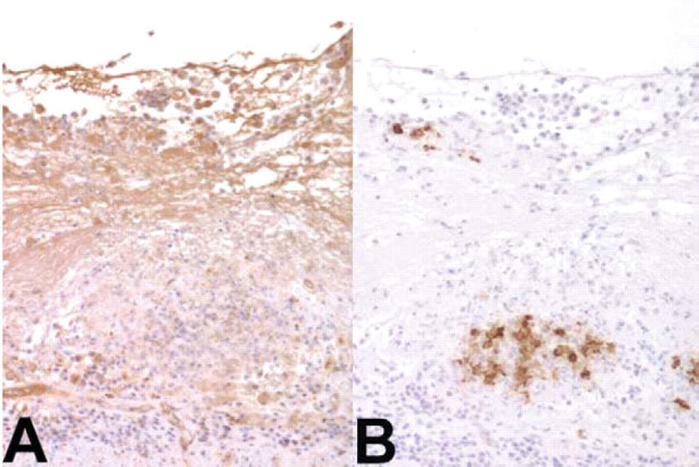
Figure 4.
Intestine. A: The fAGPrP positivity is detectable as a diffuse brown staining in perivisceral fibrin; B: FCoV antigen (brown color) is detectable within macrophage-like cells in pyogranulomatous foci. Immunohistochemistry, antibodies against human AGP (A) and FCoV (B), Mayer's hematoxylin counterstain, 100×.

