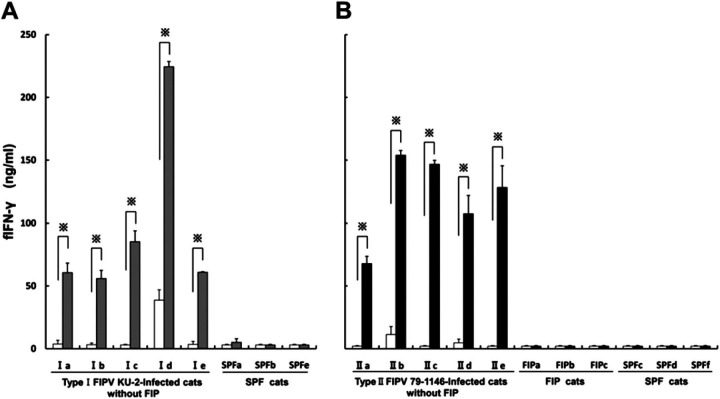
Fig 2.
Measurement of the concentration of fIFN-γ in the PBMCs culture supernatants using sandwich ELISA. PBMCs were derived from 10 FIPV-infected cats without FIP (Ia–Ie and IIa–IIe were inoculated with the type I FIPV KU-2 and type II FIPV 79-1146 strains, respectively), three FIP cats (FIPa–FIPc) and six SPF cats (SPFa–SPFf). The cells were cultured with the heat-inactivated FIPV KU-2 strain (grey bar; A), FIPV 79-1146 strain (solid bar; B), or culture medium alone (open bar). Each experiment was performed in quadruplicate. The results are expressed as means±SEM. ∗ indicates a significant difference by the t-test (P<0.01).

