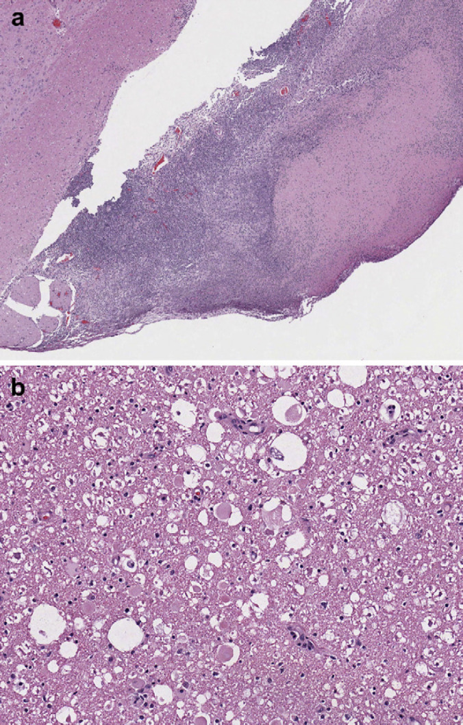
Fig 2.

(a) Section of brainstem at 4× showing large scale lymphoplasmacytic infiltration into the superficial parenchyma of the brainstem. (b) Section of brainstem at 40× showing intact and degenerative neutrophils and lymphoplasmacytic infiltration representative of severe inflammation.
