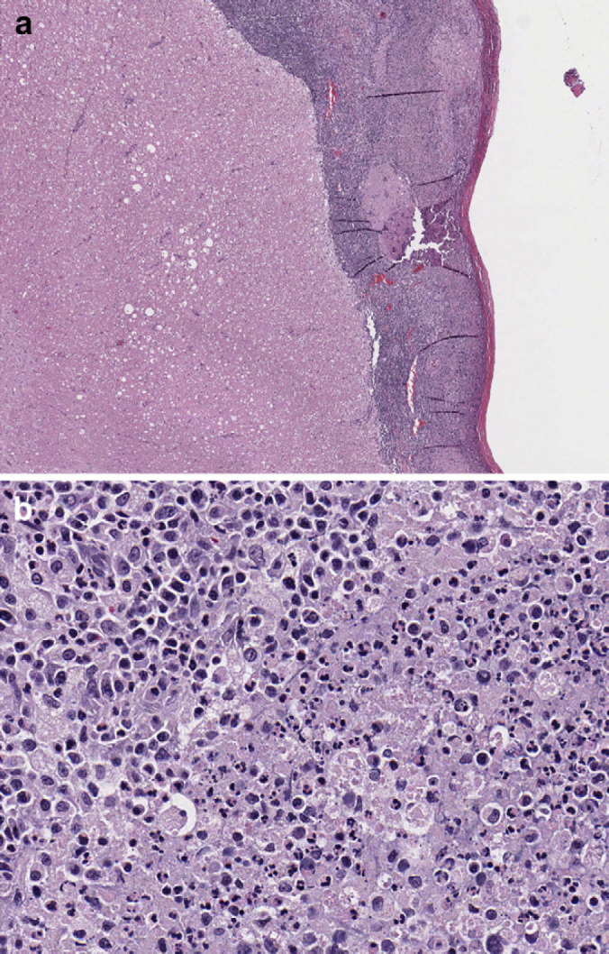
Fig 3.

(a) Section of spinal cord at 4× representative of leptomeninges from C3–C6. Multifocal randomly distributed areas of white matter contained dilated myelin sheaths with occasional dilated axons and axonophagia. Areas of parenchymal rarefaction were present in the white matter, interpreted as pan-necrosis. (b) Section of spinal cord at 40× showing leptomeninges that are markedly expanded and effaced by accumulations of intact and degenerate neutrophils admixed with eosinophilic slightly fibrillar material (fibrin). Numerous coalescing aggregates of lymphocytes, plasma cells and histiocytes were also present adjacent to the outer edge of the white matter.
