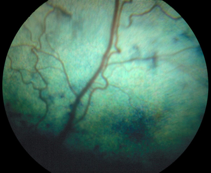
Fig 1.

Photograph of the retina showing fundic lesions associated with a chorioretinitis. Retinal arterioles demonstrate increased tortuosity and irregularity. There are areas of abnormal choroidal pigmentation with a few bullae. Numerous bullae in addition to hyperreflectivity were present in other areas of the tapetal fundus.
