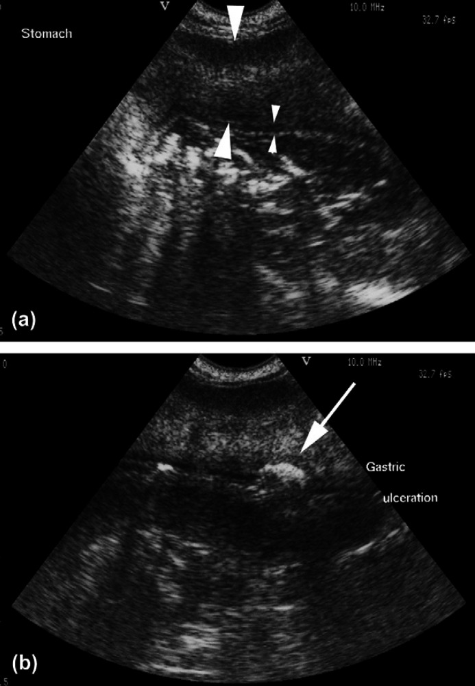
Fig 2.
Transverse (a) and oblique (b) ultrasound images of the stomach. There is massive thickening and increased echogenicity of the muscularis layer (large arrowheads) resulting in loss of visualisation of the submucosal and serosal layers. The mucosal layer is normal (small arrowheads) and clearly defined except for focal cratering (arrow) (b) suggestive of ulceration.

