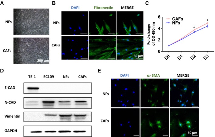
Figure 1.

Isolation and identification of cancer‐associated fibroblasts (CAFs) and normal fibroblasts (NFs). A and B, The cell morphology of CAFs and NFs was observed; grew as a fibroblast‐like, spindle‐shaped morphology of cells were represented under light microscope or Confocal microscopy. C, Cell growth assay of CAFs and NFs. D, Western blot analyses of the expression of mesenchymal marker, N‐cadherin, vimentin and epithelial and endothelial marker, E‐cadherin in those stromal cells. E, IF assay of the expression of myofibroblast marker α‐SMA in NFs and CAFs
