Figure 3.

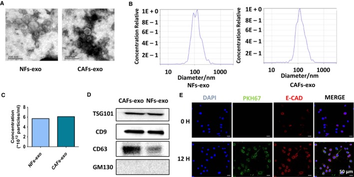
Isolation, characterization, and internalization of exosomes derived from primary stromal fibroblasts. A, Transmission electron microscopy images of cancer‐associated fibroblasts (CAFs)‐derived exosomes and normal fibroblasts (NFs)‐derived exosomes. B and C, The size and concentration of exosomes derived from CAFs and NFs examined by NTA. D, Western blot analyses of exosomal positive and negative markers (CD63, CD9, CD81, and GM130). E, Exosomes up‐taken experiment. The CAFs‐derived exosomes were labeled by PKH67 and incubated with TE‐1 cells and the green fluorescent protein‐tagged exosomes were taken up by the recipient cells after 12 h
