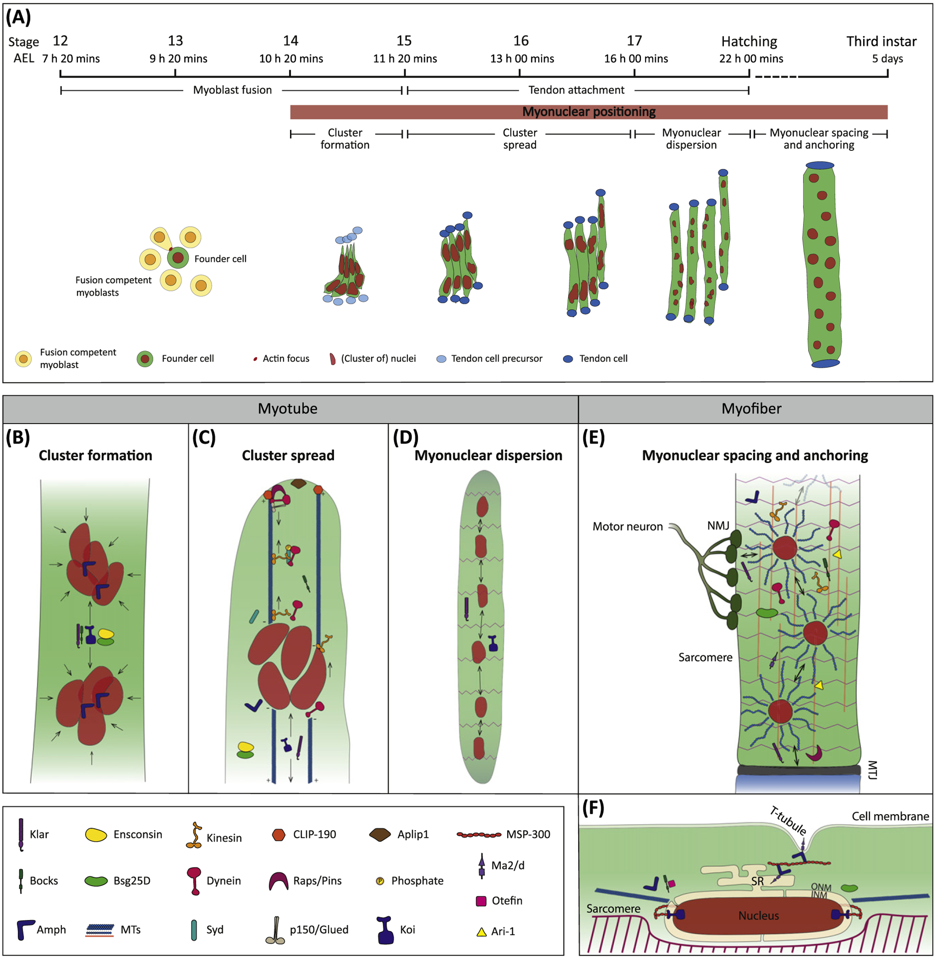
Figure 2. Schematic Representation of Myonuclear Positioning during Embryonic and Larval Drosophila melanogaster Development.

(A) Drosophila myogenesis consists of multiple steps, including myoblast fusion, tendon attachment, and myonuclear positioning. The latter is further divided into cluster formation (B), cluster spread (C), myonuclear dispersion (D), and myonuclear spacing and anchoring (E and F). Development and myonuclear positioning from stages 12 to the end of stage 17 are shown using the embryonic lateral transverse (LT) muscles as examples. From hatching to third instar larva, the muscle represented is a larval ventral longitudinal (VL) muscle. Stage and times correspond to those observed at 25°C. (B) Close-up of the two clusters of myonuclei present in the embryonic LT muscle. Arrows indicate the forces required to maintain the integrity of the two clusters and those to keep them separated. (C) Close-up of a single cluster within an embryonic LT muscle as it moves towards the end of the myotube. Arrows indicate the pulling and pushing forces clustered myonuclei experience as they move. (D) View of a single embryonic LT muscle showing the presence of sarcomeres, which, together with the neuromuscular junction (NMJ) (not shown), indicate that the muscle can experience coordinated contraction. In this panel, the double arrows represent the movements of myonuclei to be equidistant from each other. (E) View of a larval VL myofiber and its interaction with two cell types: motor neuron (dark green), forming the NMJ, and tendon (bottom of the muscle, blue), forming the myotendinous junction (MTJ). The two populations of microtubules (MTs) can be observed: longitudinal MTs run along the length of the myofiber (orange lines) and astral MTs surround the nucleus and extend in multiple directions (blue). The double arrows indicate the distance between myonuclei and the edges of the myofiber. (F) Longitudinal cross-section of a larval VL nucleus for a more detailed view of the localization of proteins involved in myonuclear spacing and anchoring. Note: protein localization in panels B-F represents the function in specific processes or mechanisms, rather than the precise described localization of each protein in the cell. Abbreviations: AEL, after egg laying; INM, inner nuclear membrane; ONM, outer nuclear membrane; SR, sarcoplasmic reticulum.
