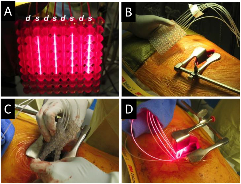
Fig. 2.
OSA for swine studies. The OSA for intraoperative PDT (IO-PDT) with four RD50 CDFs attached to a 4-channel laser diode emitting 665-nm light and four IP85 isotropic light detectors attached to our real-time dosimetry system. CDF light sources (labeled s) were spaced 20 mm apart, with isotropic detectors (labeled d) sutured to alternating channels (A). OSA before insertion into the animal (B). Flexible OSA folded for insertion through the left lateral thoracotomy incision (C). Working OSA following placement between the lung and pleural surface (D).

