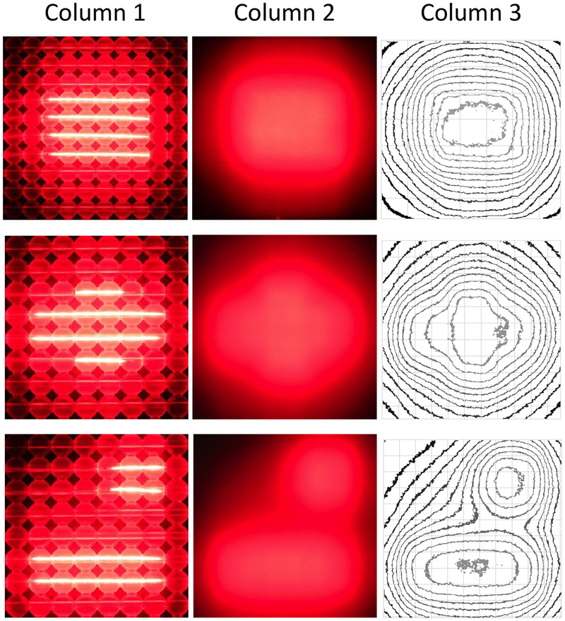
Fig. 3.
High-resolution digital images of a 10 cm × 10 cm OSA using a variety of CDF lengths and positions to provide custom shaping of the light beam. Light from the OSA alone (column 1) and through a 5-mm thick solid tissue-mimicking optical phantoms (column 2). Luminance isointensity curves were plotted using ImageJ software (column 3).

