
Figure 1.
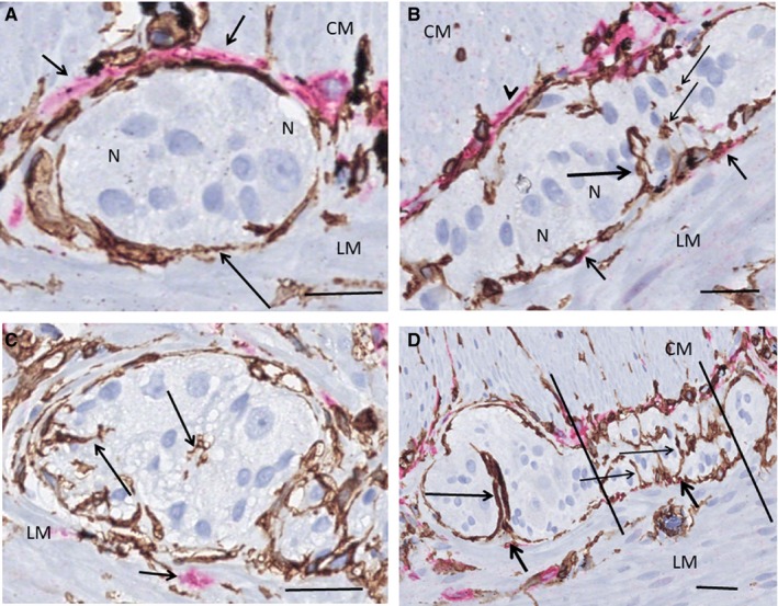
Ileum. Myenteric ganglia. Double labelling, CD34/c‐Kit; telocytes (TC): brown, interstitial cells of Cajal (ICC): red. A, TCs are in close contact with the ganglion forming a continuous layer (long arrow). The ICCs are situated between the TCs and the circular muscle (short arrows). No ICCs are observed towards the longitudinal muscle and no telopodes within the ganglion. B, Double layer of TCs and ICCs towards the circular muscle (arrowhead). Projections of two ICCs towards the longitudinal muscle (short arrows) and a telopode entering the ganglion (long arrow), probably near the origin of the interganglionic fascicle. A few transversally sectioned telopodes are also observed (thin arrows). C, Probable terminal part of a ganglion with telopodes (long arrows). There is ICC present outside the TC layer (short arrow). D, Terminal part of a ganglion with the beginning of the interganglionic nerve bundle (between the lines). There are telopodes of TCs (thin arrows) and only nuclei of glial cells are within this area. A telopode forms a semi‐septum within the ganglion (long arrow). Note a few scattered ICC projections among TCs towards the longitudinal muscle layer (short arrows). CM, circular muscle layer; LM, longitudinal muscle layer; N, neuron. Scale bars: 25 µm on actual figures