
Figure 3.
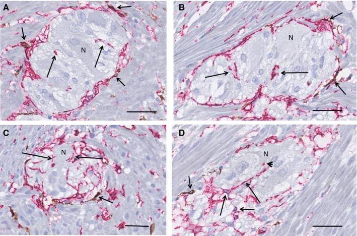
Colon. Myenteric ganglia, surrounded by longitudinal muscle cells (transversal section). Double labelling, c‐Kit/CD34; telocytes (TC): red, interstitial cells of Cajal (ICC): brown. A, An almost complete layer of TCs surrounds the ganglion with only a few scattered ICCs (short arrows). Short segments of telopodes are within the ganglion (long arrows), some are at close contact with neuron. B, The location of the few ICCs around the ganglion (short arrows) is similar to that of the ileum, that is between the telopodes and the smooth muscle cells. There are telopodes within the ganglion (long arrows). C, Telopodes are in close contact with neurons along their plasma membrane (long arrows). Short arrow shows ICCs. D, Several telopodes (long arrows) are within the ganglion even with contact with one of the neurons (arrowhead). Short arrows point to ICCs. N, neuron. Scale bars: 25 µm on actual figures