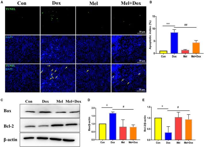
Figure 2.

Mel alleviated Dox‐induced apoptosis in H9c2 cells. Representative images of TUNEL staining (A) and quantification of apoptosis (B) in control, doxorubicin (Dox), melatonin (Mel) and Mel with Dox co‐treated H9c2 cells. Representative Western blot images (C), quantification of Bax (D) and Bcl‐2 (E) in the 4 groups of treated cells. β‐actin was used as a house‐keeping protein. n = 3 independent experiments/group. *P < .05 compared with the control group, **P < .01 compared with the control group, # P < .05 compared with the Dox‐treated group, ## P < .05 compared with the Dox‐treated group
