Figure 1.

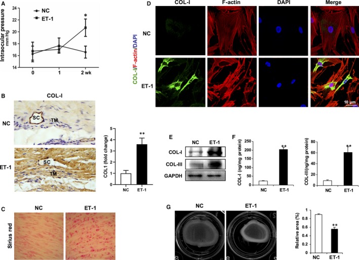
ET‐1 exposure mimics pathologic features of POAG. A, Elevated intraocular pressure (IOP) in ET‐1‐treated rabbit eyes. Topical application of ET‐1 (2 μmol/L) or PBS eye drops onto rabbit eyes 3 times a day for 2 wk, and IOPs were recorded weekly. Data are means ± SEM. n = 10 eyes per group, *P < .05 vs eyes applied with PBS (NC). B, Representative immunohistochemistry for collagen I (COL‐I) in sections of rabbit trabecular meshwork tissues. TM, trabecular meshwork; SC, Schlemm's canal. C, Deposition of collagen in cultured human trabecular meshwork cells (HTMCs) treated with ET‐1 (100 nmol/L) or PBS for 24 h were determined by Sirius Red staining. D, Representative immunofluorescence for collagen I (COL‐I) (green) or F‐actin (red). Nuclei were counterstained with DAPI (blue). Scale bar, 10 μm. E, Deposition collagen in cultured HTMCs were determined by Western blotting. F, Secretion of collagen in cultured HTMCs were determined by ELISA. Data are means ± SEM of three independent experiments. **P < .01 vs PBS control (NC). G, The contractility of HTMCs was determined by a collagen gel contraction assay. Representative images are shown. The area of the collagen disc was measured using Image J. The contraction degree was expressed as a percentage of the 24‐well area. The cultured HTMCs are from one human donor. Data are means ± SEM of three independent experiments. **P < .01 vs control
