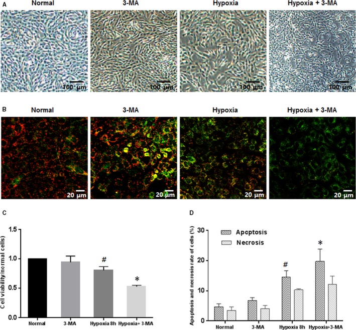
Figure 3.

Inhibiting autophagy under hypoxic conditions increased apoptosis and decreased cell viability. (A) The morphologies of 661w cells treated with 3‐MA under hypoxia altered obviously, while those exposed to 3‐MA under normoxic condition did not. Magnification: 4×. (b) ΔΨm analyses by JC‐1 staining. The ΔΨm in cells cultured in hypoxia was diminished, as revealed by increased green fluorescence, and adding 3‐MA into cultures under hypoxic conditions further exacerbate the ΔΨm alteration. Magnification: 20×. (C and D) When autophagy was inhibited in 661w cells under hypoxic conditions, cell viability was decreased while apoptosis was increased. #: P < .05, compared to normal and 3‐MA cells, *: P < .05, compared to hypoxic cells. These assays were repeated for three times
