
Figure 5.
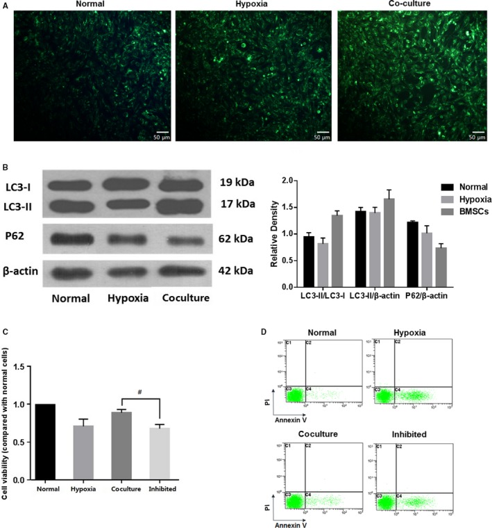
Coculturing with BMSCs increased autophagy in 661w cells, and the effect of BMSCs was weakened when autophagy was inhibited. (A) MDC staining showed higher number of autophagosome in hypoxic cells, as compared to normal cells, and the autophagosome was further increased in the cocultured group. Magnification: 20×. (B) In the cocultured group, the expression of LC3II and the ratio of LC3II/LC3I were obviously increased, while the expression of p62 was decreased, compared with those in the hypoxia and normal groups. (C) 3‐MA, an autophagy inhibitor strongly diminished the protective effects of BMSCs. In the autophagy‐inhibited coculture (inhibited) group, the cell viability was decreased to nearly that in the hypoxia group. These assays were repeated for three times. *: P < .05, compared with the other two groups. #: P < .05, compared with cocultured cells