Figure 6.

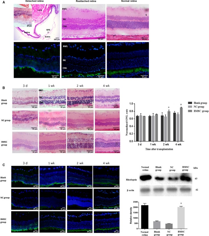
BMSC transplantation ameliorated cell death during retinal detachment. (A) In the detached retina, decreased ONL thickness, shortened photoreceptor outer segments and disordered arrangement were observed after 1 d of RD. Even after the retina was reattached, damage was still observable, as the whole retinal layer was thinned and undulated, especially in the ONL, and scattered nuclei were observed. Compared to normal retina, detached and reattached retinas were disordered and showed decreased expression of rhodopsin, as revealed by weak green fluorescence. Magnification: the photograph of detached retina from HE staining was 4×, while others (including inset) were 20×. (B) Histological changes were ameliorated in the BMSC‐transplanted group, as a thicker ONL was observed, especially at the 2 and 4 wk after transplantation. Magnification: 20×. (C) Photoreceptors were more regularly arranged and thicker in BMSC‐transplanted groups, as detected by immunohistochemistry (magnification: 40×), with rhodopsin expression being increased to nearly normal levels after the retina was reattached. These assays were repeated for three times. *: P < .05, compared with the blank and NC groups
