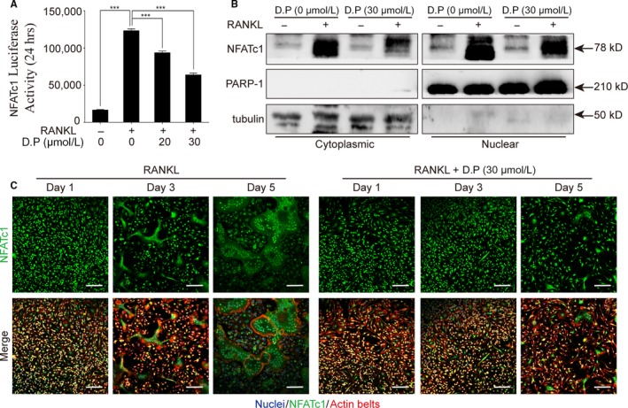
Figure 7.

D.P inhibited NFATc1 activity and translocation to nuclei. (A) NFATc1 activity was detected using a luciferase reporter gene assay. n = 3; ***P < .001. (B) Representative images of Western blot to determine NFATc1 translocation after stimulation with RANKL for 5 days in the presence or absence of D.P. PARP‐1 and tubulin were used as cytoplasmic and nuclear loading controls, respectively. (C) Representative images of the immunofluorescent microscopy co‐stained for NFATc1 (green), actin belts (red) and nuclei (blue) (Scale bar = 100 μm)
