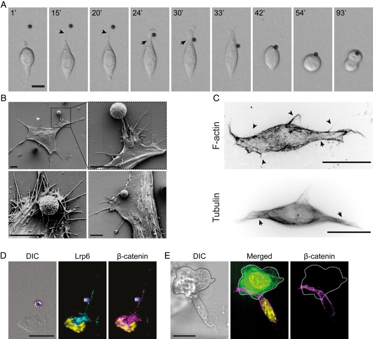
Fig. 2.
ESCs extend exploratory actin-based cytonemes that recruit localized Wnt signals to the plasma membrane. (A) Representative frames from time-lapse imaging of an ESC contacting a Wnt3a bead (black sphere) with a cytoneme. Time is expressed in minutes. (Scale bar, 10 μm.) Arrowheads indicate thin cytonemes; arrows indicate larger cytonemes used for Wnt-bead recruitment. (B) Scanning electron microscopy images of ESCs at various stages of interaction with the Wnt3a bead (white/gray spheres). White arrowhead indicates a thin cytoneme. (Scale bar, 5 μm.) (C) Deconvolved structural illumination images of ESCs stained with the live-cell cytoskeleton-staining reagents SiR-Actin (Top) and SiR-Tubulin (Bottom). Arrows indicate larger cytonemes; arrowheads indicate thin cytonemes. (Scale bars, 10 μm.) (D) Representative images of an ESC cytoneme contacting a Wnt3a bead (dashed purple circle or 3D-reconstructed purple sphere) stained with antibodies against LRP6 (cyan) and β-catenin (magenta) with DAPI (yellow). (Scale bar, 20 μm.) (E) Representative images of an ESC contacting an eGFP-expressing TSC (green, dashed edge) stained with antibodies against β-catenin (magenta) with DAPI (yellow). (Scale bar, 20 μm.)

