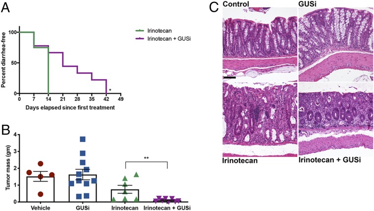
Fig. 4.
GUSi improves irinotecan efficacy in C3TAg breast cancer GEMM. (A) Kaplan–Meier analysis of remaining diarrhea free with GUSi cotreatment with irinotecan. P < 0.05 by log-rank (Mantel–Cox) test. (B) Irinotecan reduces tumor masses in C3Tag animals, while IRI + GUSi significantly diminishes tumor masses compared with irinotecan alone. **P < 0.01 by one-way ANOVA with Sidak’s multiple comparisons test. (C) Representative sections of colon revealing no significant microscopic differences between untreated control (Upper Left) and GUSi-treated (Upper Right) mice. Colons of IRI-treated mice (Lower Left) display moderate inflammation and epithelial damage, with the lamina propria expanded by inflammatory cells (arrows), variably sized and irregularly shaped crypts lined by attenuated epithelial cells with lumens containing sloughed epithelial cells and inflammatory cells (filled circles), and a fragmented and irregular overlying mucosal epithelium (filled square). While the colons of IRI + GUSi-treated mice (Lower Right) display inflammation in the lamina propria (arrow) and a few remaining damaged crypts at the superficial surface (filled circle), there is abundant crypt regeneration as the deeper mucosa contains a line of regenerative crypts lined by plump, elongated cells with cytoplasmic basophilia (asterisks) and the overlying epithelium displaying outstretched epithelial cells in the process of repair (open square). (Scale bar, 100 μM.)

