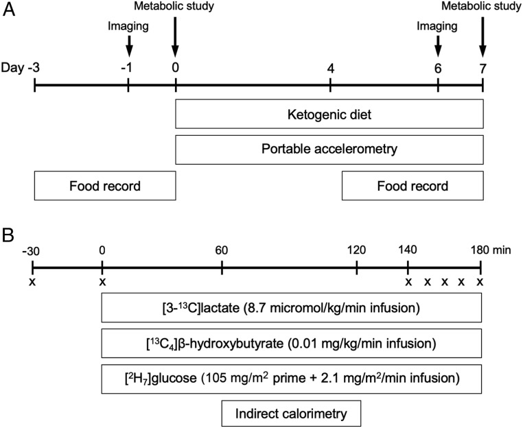
Fig. 1.
Study design. (A) Before and after the 6-d KD, participants visited an imaging center for measurement of IHTG content and liver stiffness (days −1 and 6) and underwent metabolic studies at the Clinical Research Unit (days 0 and 7). Participants wore portable accelerometers between days 0 and 7 for determination of physical activity and recorded 3-d food intake starting at days −3 and 4 for determination of dietary composition and compliance. (B) During metabolic study visits, 180-min tracer infusions of lactate, β-OHB, and glucose were given for determination of rates of substrate fluxes. Indirect calorimetry was performed to measure energy expenditure and rates of substrate oxidation. An “X” denotes blood sample.

