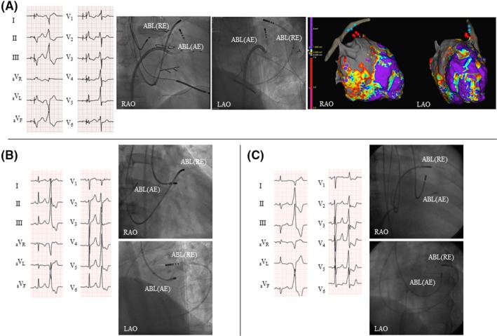
Figure 1.

12‐lead electrocardiograms (ECGs) and fluoroscopic views for the 3 cases. A, Case 1. The 12‐lead ECG shows biventricular pacing (the first beat) and ventricular premature complex (VPC, the second beat) with the right bundle branch block and inferior axis. Fluoroscopic views show the catheters during bipolar ablation between the left ventricular (LV) endocardium (active electrode (AE); irrigated catheter) and great cardiac vein (GCV, return electrode (RE); an 8‐mm‐tip catheter). EnSite images of the left ventricle and coronary sinus show the ablation sites. The red tags are bipolar ablation sites, and the blue tags are unipolar ablation sites from the GCV. B, Case 2. The 12‐lead ECG shows the sinus rhythm (the first beat) and VPC (the second beat) with the left bundle branch block and inferior axis. Fluoroscopic views show the catheters during bipolar ablation between the LV endocardium (AE, irrigated catheter) and anterior interventricular vein (GCV, RE, 8‐mm‐tip catheter). C, Case 3. The 12‐lead ECG shows the sinus rhythm (the first beat) and VPC (the second beat) with the right bundle branch block and inferior axis. Fluoroscopic views show the catheters during bipolar ablation between the LV endocardium (AE, irrigated catheter) and GCV (RE, 8‐mm‐tip catheter). ABL, ablation catheter; AE, active electrode; LAO, left anterior oblique; RAO, right anterior oblique; RE, return electrode
