-
A–D
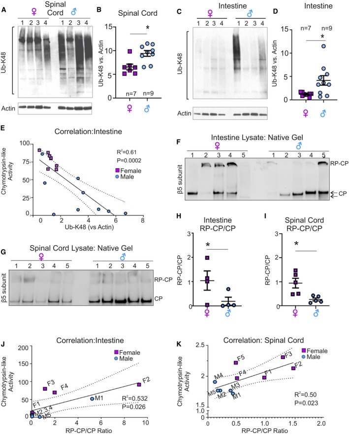
Western blot analysis for Ub‐K48‐linked proteins in spinal cord (A) and intestine (C) lysates from male and female mice. (B, D) Quantification of panels (A) and (C), respectively. Values were normalized to actin as a loading control. Each central line and error bar indicate mean ± SEM of a biological replicates.
-
E
Curve of correlation between chymotrypsin‐like activity and Ub‐K48‐linked proteins in the intestine, where variability between mice is observed.
-
F
Native gel followed by Western blot of β5 subunit to distinguish unassembled catalytic core (CP) or assembled with regulatory core (RP) in male (n = 5) and female (n = 5) mice in intestine.
-
G
Native gel followed by Western blot of β5 subunit to distinguish unassembled catalytic core (CP) or assembled with regulatory core (RP) in male (n = 5) and female (n = 5) spinal cord.
-
H
Quantification of the ratio of assembled (RP‐CP) versus unassembled (CP) proteasome in the intestine of males and females. Each central line and error bar indicate mean ± SEM of a biological replicates (n = 5 per group).
-
I
Quantification of the ratio of RP‐CP versus CP proteasome in the spinal cord of males and females. Each central line and error bar indicate mean ± SEM of a biological replicates (n = 5 per group).
-
J
Correlation curve between chymotrypsin‐like activity and RP‐CP/CP ratio in individual female (purple) and male (blue) intestine.
-
K
Correlation curve between chymotrypsin‐like activity and RP‐CP/CP ratio in individual female (purple) and male (blue) spinal cord.
‐test comparing male and female samples.

