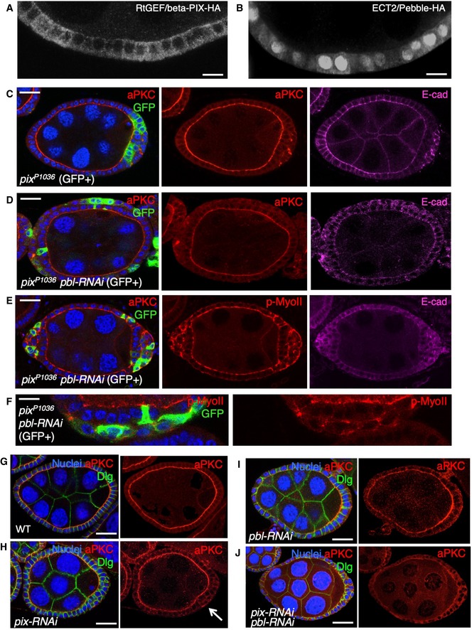
Cross‐section of cuboidal follicular epithelium showing cytoplasmic localisation of overexpressed Cdc42 GEF beta‐PIX tagged with HA. Scale bar ˜3 μm. n > 4 independent biological replicates.
Cross‐section of cuboidal follicular epithelium showing nuclear and cytoplasmic localisation of overexpressed ECT2/Pbl tagged with HA. Scale bar ˜3 μm. n > 10 independent biological replicates.
Stage 7 egg chamber in which follicle cells have mutant clones for pix
P1036, marked by expression of GFP, shows extrusion of cells which retain abnormal aPKC and E‐cad localisation. Scale bar ˜6 μm. n > 4 independent biological replicates.
Stage 7 egg chamber in which follicle cells have mutant clones for pix
P1036 also expressing UAS.Pbl‐RNAi, marked by expression of GFP, shows extrusion of cells featuring loss of aPKC plasma membrane localisation, along with abnormal E‐cad localisation. Scale bar ˜6 μm. n > 3 independent biological replicates.
Stage 7 egg chamber in which follicle cells have mutant clones for pix
P1036 also expressing UAS.Pbl‐RNAi, marked by expression of GFP, shows extrusion of cells featuring mislocalisation of p‐MyoII (p‐MLC), along with abnormal E‐cad localisation. Scale bar ˜6 μm. n > 3 independent biological replicates.
High‐magnification view of mutant clones for pix
P1036 also expressing UAS.Pbl‐RNAi, marked by expression of GFP, shows extrusion of cells featuring mislocalisation of p‐MyoII (p‐MLC). Scale bar ˜3 μm. n > 3 independent biological replicates.
Wild‐type stage 7 egg chamber in which follicle cells have normal aPKC and Dlg localisation. Scale bar ˜6 μm. n > 4 independent biological replicates.
tj.Gal4‐driven UAS.pix‐RNAi causes mild extrusion of cells with abnormal aPKC localisation (arrow). Scale bar ˜6 μm. n > 5 independent biological replicates.
tj.Gal4‐driven UAS.pbl‐RNAi causes enlarged cells with normal morphology and aPKC localisation. Scale bar ˜6 μm. n > 3 independent biological replicates.
tj.Gal4‐driven UAS.pix‐RNAi and UAS.pbl‐RNAi causes loss of aPKC throughout the follicular epithelium, while Dlg spreads ectopically. Scale bar ˜6 μm. n > 9 independent biological replicates.