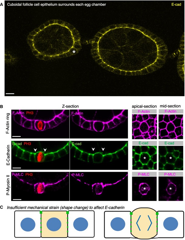
Figure 8. The cuboidal follicle cell epithelium does not downregulate E‐cadherin at mitosis.

- Early stage Drosophila egg chambers showing giant germ‐line nurse cells surrounded by E‐cadherin‐positive follicle cell epithelium. A follicle cell undergoing mitotic rounding is highlighted with an asterisk (*). Scale bar ˜3 μm. n > 3 independent biological replicates.
- High‐resolution imaging of the F‐actin cortex, E‐cadherin and phospho‐MyoII in a mitotically rounded follicle cell. Note maintenance of adherens junctions including both E‐cad and the contractile actomyosin ring during mitotic rounding. Asterisks or pH3 staining indicates a single mitotic cell. Scale bar ˜3 μm. n > 4 independent biological replicates.
- Schematic diagram of follicle cell rounding, showing the relatively mild shape change that is insufficient to affect E‐cadherin levels during mitotic rounding.
