
Figure EV5. YTHDF1 regulates the translation of TCF7L2 during Wnt activation.
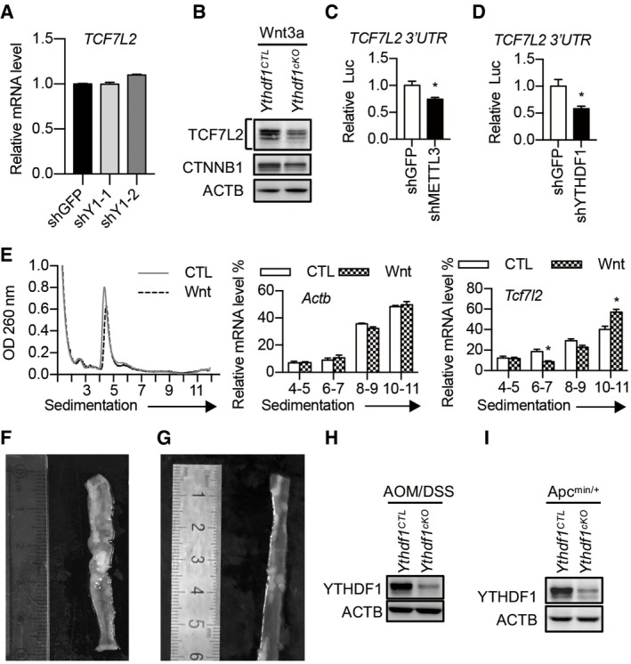
- TCF7L2 mRNA level in control and YTHDF1 knockdown HCT116 cells. Data are represented as mean ± SEM. 3 biological replicates.
- Immunoblot analysis of intestinal crypts from WT or Ythdf1 cKO mice treated with Wnt3a (60 ng/ml) for 30 min.
- Dual‐Luciferase Assay with a construct bearing the 3′UTR of TCF7L2 in HCT116 cells with or without METTL3 knockdown. Data are represented as mean ± SEM. *P < 0.05 (3 biological replicates, t‐test).
- Dual‐Luciferase Assay with a construct bearing the 3′UTR of TCF7L2 in HCT116 cells with or without YTHDF1 knockdown. Data are represented as mean ± SEM. *P < 0.05 (3 biological replicates, t‐test).
- Polysome profiles of mouse intestinal crypt treated with or without Wnt3a treatment for 30 min. The left panel is the same experiment as Fig 1B. The right panels show the distributions of Tcf7l2 and Actb in polysome fractions. Data are represented as mean ± SEM. *P < 0.05 (3 biological replicates, t‐test).
- Mouse colon after 6 weeks of AOM/DSS induction.
- Small intestine from a 3‐month‐old Apc min/+ mouse.
- Western blot showing YTHDF1 knockout efficiency in AOM/DSS‐induced tumors.
- Western blot showing YTHDF1 knockout efficiency in Apc min/+ tumors.
Source data are available online for this figure.