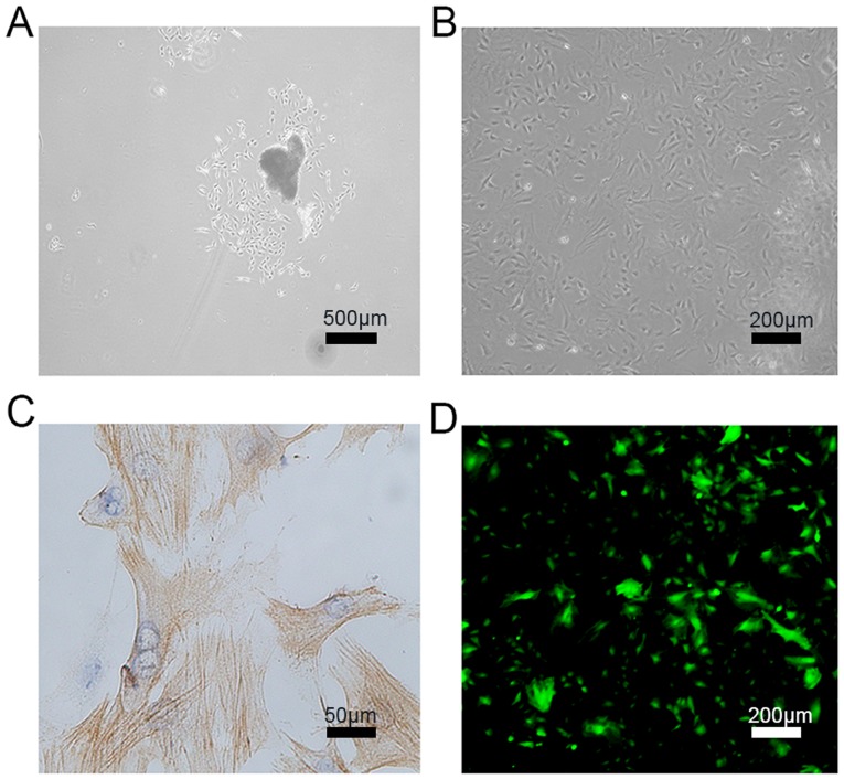
Figure 3.
Characterization of primary PASMCs isolated from pulmonary arteries of rats. (A) PASMCs isolated from the pulmonary artery mass and aggregated into clusters (5 days after start of culture). (B) PASMCs exhibiting long fusiform shapes and ‘peak-valley’ appearance (10 days after start of culture). (C) Immunocytochemistry staining confirmed α-smooth muscle actin expression based on the specific myofilament structure of PASMCs (15 days after start of culture). (D) Specific TMEM16A-siRNA lentiviral vectors, which also encode GFP, were transfected into PASMCs and observed under a light microscope. PASMCs, pulmonary artery smooth muscle cells; TMEM16A, transmembrane member 16A; siRNA, small interfering RNA.

