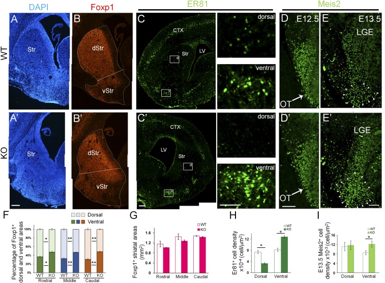
Fig. 1.
Abnormal structural alteration in the developing striatum of Nolz-1 KO brains. (A and A') DAPI staining shows the phenotype of the shrunk dorsal striatum and expanded OT of the ventral striatum in E18.5 Nolz-1 KO brains. (B and B') Foxp1+ dorsal and ventral striata, respectively, are decreased and increased in E18.5 Nolz-1 KO brains. (C and C') Larger Er81+ cell clusters are present in the OT of the E18.5 Nolz-1 KO brain compared to WT. Scattered Er81+ cells are nearly absent in the dorsal KO striatum. The boxed regions in D and D′ are shown at high magnification. (D and D') At E12.5, the Meis2 expression pattern in the the LGE is similar between WT and Nolz-1 KO brains. (E and E') By E13.5, the Meis2+ region is increased in the ventral part of the KO LGE. (F) Quantification shows decreases and increases, respectively, in Foxp1+ dorsal and ventral striata of E18.5 Nolz-1 KO brains. (G) The total area of the striatum is not changed in the Nolz-1 KO striatum from R to C levels. (H) Er81+ cells are decreased and increased, respectively, in the dorsal and ventral KO striata. (I) Meis2+ cells are increased in the ventral part of the KO LGE. *P < 0.05; **P < 0.01; n = 3/group. (Scale bars, 200 μm [A–C′], 100 μm [C and C′] for high magnification, 100 μm [D and E′].) Photomicrographs in A–C′ are stitched images.

